


For a cow to conceive successfully, the quality of follicle development is key. Veterinary ultrasound machines have now become a “helping hand” on farms, allowing us to directly observe the entire process of a follicle growing from a tiny “dot” to full maturity. Through the ultrasound images displayed on the screen, farmers can easily determine the best breeding time for cows—saving time and improving conception rates. Today, let’s talk about how cow follicles develop step by step, and how ultrasound can make breeding programs more accurate.
Five Key Stages of Follicle Development
Follicle development in cows is like building a house—it must be done layer by layer. It starts with the primordial follicle, where follicle cells are quietly lined up flat in the ovary, like bricks lying on the ground. Then comes the primary follicle stage, where the cells start to become three-dimensional, stacking into multiple layers, like bricks forming small columns. At this point, an ultrasound can already show some signs—although the follicle is less than 5 mm in diameter, the screen will display a small dark area, which is the follicle in its early form.
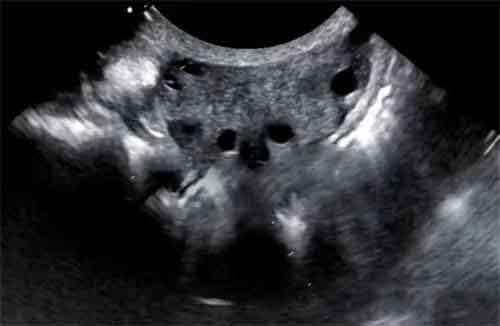
In the secondary follicle stage, a transparent zona pellucida forms around the follicle, like a protective shell. On ultrasound, the follicle diameter increases to about 5–8 mm, and the shape becomes rounder. When the follicle enters the tertiary follicle stage, fluid begins to accumulate inside, forming a small cavity. The diameter grows to about 8–12 mm, and the dark area on ultrasound becomes more pronounced with clearer edges.
Finally, the mature follicle stage arrives. The follicle reaches 15–20 mm in diameter, like a water-filled balloon. On ultrasound, the follicle wall appears thinner, with most of the interior showing a dark fluid area. At this time, cows also show obvious estrus signs—such as vulva swelling or mounting other cows—signals that the body is saying: “I’m ready to breed!”
Using Veterinary Ultrasound to Pinpoint the Best Breeding Time
For successful breeding, knowing follicle size is not enough—you must also capture the moment of ovulation. Continuous ultrasound monitoring shows that a mature follicle will suddenly “disappear” about 12 hours after estrus—that’s ovulation. For example, if you see an 18 mm follicle on ultrasound in the morning and the cow is still in heat, the best time for insemination is later that afternoon or the next morning.
Some farmers may notice that a follicle stops growing after reaching 10 mm. In this case, don’t rush to breed. If ultrasound shows no change for three consecutive days, it may indicate arrested development, requiring feed adjustment or a health check. On the other hand, if a follicle grows too quickly—for instance, jumping from 8 mm to 15 mm in just two days—it could be due to hormonal imbalance, which also lowers conception rates.
How Ultrasound Helps Avoid Pitfalls
In the past, rectal palpation was the only way to judge follicles, relying solely on experience and touch. Now, ultrasound provides clear images, giving farmers confidence. For example, it can also distinguish between a normal corpus luteum and a cyst. On ultrasound, a normal corpus luteum has neat edges and a uniform interior. But if you see dark fluid-filled areas inside, it’s likely a cyst, which needs to be treated promptly.
Post time: Aug-19-2025