


Quick answer
For equine imaging, a 17x17-inch flat panel detector is usually the stronger choice because it delivers broader anatomy coverage, fewer positioning compromises, and smoother imaging in demanding field or room-based scenarios. For small animal imaging, 14x17-inch DR detectors remain a practical all-purpose option for routine canine and feline studies, while 17x17 is often the better fit for hospitals that want wider coverage, fewer repeat positions, and more flexibility for larger dogs and orthopedic work. 10x12 detectors still have a place, but they are usually more specialized than universal.
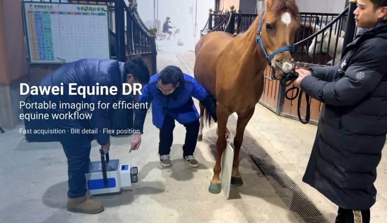
This size logic is reflected in current veterinary DR configurations. Dawei RV-80E, built for large-animal applications, uses a 43 cm x 43 cm (17x17-inch) detector with a 3072 x 3072 matrix, 139 μm pixel size, and 16-bit conversion, which fits the imaging demands of larger anatomy and higher exposure requirements.
Why detector size matters in veterinary DR
When evaluating a veterinary DR system, detector size shapes anatomy coverage, positioning efficiency, repeat exposure risk, handling comfort, exam speed, and overall workflow consistency.
Choosing the right flat panel detector size is not just about fitting anatomy on the screen. It also affects how efficiently a clinic can work every day.
DVM360 notes that the active imaging area is usually slightly smaller than the detector’s outside dimensions, which is especially relevant in veterinary radiography. In practical system design, active area often matters more than nominal size alone. Dawei RV-32AL is positioned in the 17x17 class, while its listed active imaging area is 404 mm x 404 mm. Dawei RV-5E lists an effective area of 427 mm x 427 mm, and Dawei RV-32A and Dawei RV-32B list 430 mm x 430 mm imaging area. These differences show why usable field of view matters as much as the detector label.
The detector sizes most veterinary buyers compare
Most veterinary DR purchasing decisions still revolve around three familiar size categories:
| Nominal size | Approx. metric size | Typical veterinary use |
| 10x12 in | about 25x30 cm | Dental, exotics, extremities, compact portable workflows |
| 14x17 in | about 35x43 cm | General small animal imaging, daily canine/feline radiography |
| 17x17 in | about 43x43 cm | Equine imaging, larger dogs, orthopedic work, broad-coverage DR rooms |
A current Vieworks detector lineup continues to reflect the same core market structure, showing mainstream panels around 25x30 cm, 36x43 cm, and 43x43 cm.
Large-area detector design is also evident in several Dawei DR models. Dawei RV-32A, RV-32B, RV-32AL, RV-80E, and RV-5E all sit at the large-format end of the veterinary DR spectrum, with active imaging areas around 404 mm x 404 mm, 427 mm x 427 mm, or 430 mm x 430 mm. This makes large-format DR especially relevant for clinics that want broader application range from a single system.
Equine imaging: why 17x17 is usually the better choice
When the application is equine DR, the buying decision becomes more straightforward. Horses create larger anatomy targets, more positioning variability, and less tolerance for clipped regions of interest. In those scenarios, 17x17 is not just a larger detector. It is usually the more dependable workflow tool.
A larger panel helps capture more anatomy in a single exposure, reduces repositioning, and lowers the chance of incomplete coverage. A veterinary page from MidXray / MyVet Imaging highlights the value of 17x17 detectors for offering a fuller field of view and reducing the need to rotate the detector for larger anatomy. The same logic appears in Dawei RV-80E, which combines a 17x17 / 43 cm x 43 cm detector with 3.7 lp/mm spatial resolution and 3–5 second imaging time, supporting efficient large-animal imaging.
The full system configuration points in the same direction. Dawei RV-80E also lists up to 80 kW output, 40–150 kV, and 0.1–1000 mAs, a specification package built for larger and more demanding studies. For equine practices and distributors serving the large-animal segment, a broad detector size is usually the more logical long-term match.
Small animal imaging: why 14x17 remains a strong default
In small animal DR, the workflow is more varied. Cats, small dogs, medium dogs, large breeds, abdominal studies, thorax views, orthopedic cases, and trauma work may all happen in the same room. That is why 14x17-inch flat panel detectors continue to hold a strong position. They offer a balanced combination of coverage, handling comfort, and room compatibility.
A 14x17 detector is often large enough for routine canine and feline imaging while still feeling manageable for daily operation. It can also be easier to introduce into an existing room.
At the same time, many clinics consider moving directly to 17x17, especially when they see more large dogs, want fewer repeat positions, or expect one DR room to handle a broader range of anatomy. Dawei RV-32A and Dawei RV-32B illustrate this direction well. Both use a detector with 430 mm x 430 mm effective imaging area, 3072 x 3072 pixels, 16-bit A/D conversion, and roughly 139–140 μm pixel pitch. These specifications support wider coverage and fewer compromises in daily pet imaging.
Dawei RV-32AL supports the same trend from another angle. Its 404 mm x 404 mm active imaging area, 3072 x 3072 pixel matrix, 139 μm pixel pitch, and 16-bit conversion show how a room-based animal DR system can be configured for broad clinical use rather than narrow single-purpose imaging.
Why 10x12 still has a place
Although 10x12 DR detectors are not usually the main recommendation for a general-purpose veterinary DR room, they still solve specific problems well.
They are often relevant for:
- · dental imaging,
- · exotics,
- · smaller anatomy,
- · extremities,
- · and compact portable workflows where maneuverability matters.
The broader market trend, however, continues to favor larger active areas for clinics that want one detector to cover the majority of routine DR work efficiently. That same preference can be seen in large-area Dawei configurations such as RV-32A, RV-32B, and RV-32AL, where broad detector coverage supports more flexible daily workflow.
Image quality still matters just as much as size
A larger detector does not automatically mean better diagnostic quality. Coverage is one part of performance. The rest depends on detector architecture, scintillator choice, conversion depth, generator stability, and image processing.
Several Dawei DR models show how these specifications are combined in practice. Dawei RV-32AL uses amorphous silicon, CsI, 139 μm pixel pitch, and 16-bit ADC. Dawei RV-5E uses CsI:Tl, a 3072 x 3072 effective array, 3-second preview time, and 5-second imaging time. Dawei RV-80E also combines CsI, 139 μm, and 16-bit conversion. These configurations reflect a broader veterinary DR trend toward maintaining image detail while expanding usable anatomy coverage.
Larger format can support both coverage and diagnostic confidence when detector technology and overall system integration are strong.
Portable vs room-based DR
Portable DR and room-based DR are different workflow choices.
In a fixed DR room, a clinic can usually absorb a larger detector more comfortably because alignment, handling, and positioning are more controlled. In portable work, size and weight become more important for the technician using the system every day.
Dawei RV-5E shows how portable workflow does not always mean a smaller detector. Even as a portable animal DR system, it still uses a large detector format with 427 mm x 427 mm effective area, 139 μm pixel size, and 3.6 lp/mm spatial resolution. At the same time, its 5.3 kW generator shows how portability depends on the balance of the full system rather than the panel alone.
For mobile and multi-scenario imaging, larger detector coverage may still be the better fit. For compact and highly specific workflows, a smaller format may be easier to manage.
Market direction in 2026
Exact veterinary-only shipment numbers remain difficult to verify publicly, but the broader X-ray detector market is still growing. A 2026 MarketsandMarkets estimate places the wider segment at USD 3.99 billion in 2026, with a forecast of USD 5.35 billion by 2031 at a 5.0% CAGR. Veterinary imaging is only one part of that market, but the direction is clear: detector investment is continuing, and clinics are still modernizing.
Across vendor pages, product lineups, and current system configurations, the same priorities appear repeatedly:
- · faster image availability,
- · broader anatomy coverage,
- · lower dose,
- · detector durability,
- · software integration,
- · and fewer workflow interruptions.
Dawei DR configurations align closely with these priorities. Models such as RV-32AL, RV-5E, and RV-80E combine large-format detectors, 16-bit conversion, CsI-based imaging, and fast acquisition characteristics, reflecting the market shift toward workflow efficiency together with image quality.
Which detector size should a buyer choose?
If the workflow is mainly equine imaging, 17x17 is usually the most natural recommendation. It aligns with anatomy scale, field workflow, and long-term usability. Dawei RV-80E reflects that logic through its detector size, generator power, and exposure range.
If the workflow is primarily general small animal imaging, 14x17 remains a strong choice, especially for clinics that want reliable everyday performance with familiar handling. But if the clinic sees larger dogs, orthopedic cases, or wants broader one-room capability, 17x17 becomes increasingly attractive. Dawei RV-32A, RV-32B, and RV-32AL all support this direction with large-area detectors, 16-bit conversion, and 139–140 μm class pixel pitch.
If the workflow is narrower, such as dental, exotic species, or smaller dedicated studies, 10x12 still has value. It is simply less likely to be the best one-detector answer for a busy general-practice veterinary clinic.
Practical takeaway
The most effective detector choice is usually the one that removes the most workflow friction without creating unnecessary complexity.
- · If maximum flexibility across larger anatomy and more demanding studies is the priority, 17x17 is usually the better fit.
- · If the goal is a dependable all-round choice for day-to-day companion animal work, 14x17 remains highly competitive.
- · If the use case is narrower and more specialized, 10x12 can still be the right answer.
For many clinics, the more useful question is not which detector size is most popular, but which size will still fit the workflow two or three years from now.
FAQ
Is 17x17 too large for small animal imaging?
Not necessarily. Many hospitals use 17x17 successfully because it helps with larger dogs, chest studies, abdomen work, and orthopedics. The main tradeoff is handling and room fit, not whether the format is clinically useful.
Is 14x17 enough for equine imaging?
It may work for selected studies, but it is usually less forgiving. If equine imaging is a core part of the workflow, 17x17 is generally the safer investment.
Does a larger detector reduce image quality?
No, not by itself. Image quality depends on pixel pitch, scintillator type, ADC depth, generator stability, and software processing. Configurations such as Dawei RV-32AL, RV-5E, and RV-80E show how large-format systems can still preserve detail while improving coverage.
What should buyers compare besides detector size?
Buyers should compare active imaging area, pixel pitch, spatial resolution, scintillator, wired or wireless workflow, detector durability, acquisition speed, and overall system integration.
Post time: Jun-08-2026