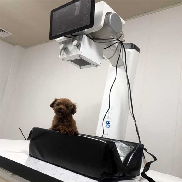
Digital Radiography (DR) is an advanced medical imaging technique used to obtain X-ray images of structures in the animal’s body and to digitize these images for diagnosis, analysis and storage. Compared with traditional film X-ray imaging, digital X-ray imaging has many advantages, including higher image quality, lower radiation dose, DR photography with its high-quality images and high efficiency is gradually replacing the traditional X-ray photography technology, and has become the representative of today’s digital X-ray photography technology.
1 The advantages of veterinary x ray
1.1 Rapid diagnosis
DR diagnosis can display the image within a few seconds after exposure. This feature has a high value in clinical applications. When there are seriously ill animals or emergency animals need to X-ray rapid diagnosis to clarify the cause of the disease, DR examination can reduce the traditional X-ray film due to the time spent on film shuffling, to win the valuable time for rescue and treatment. In addition, the short exposure time and fast acquisition speed of DR can maximize the elimination of artifacts brought about by the involuntary movement of animals.
1.2 Smaller irradiation dose for examined animals
The traditional X-ray machine applied in veterinary clinics generally has the problem of large single exposure dose and strong radiation. In the diagnostic work of certain diseases, it is often necessary to repeat the X-ray examination several times, which increases the irradiated dose of the animal. Excessive irradiation may cause turbidity of eye crystals, cataracts, hematopoietic dysfunction, reduced immune function, cancer and other diseases.DR has the advantages of small irradiation dose and less irradiation time, which makes the irradiation damage of examined animals effectively reduced.
1.3 Higher dynamic range and larger contrast range, richer image levels
In the application of DR to the skeletal and muscular system examination, in order to make the soft tissue structure appear more clearly, it can be completed by using low contrast and high brightness processing; while using high contrast and low brightness processing can determine the lesions of bone trabeculae and bone cortex. Using dual-energy subtraction technology, the bone tissue image has no soft tissue overlap, which is suitable for the examination of rib bone diseases.
1.4 High accuracy
In the X-ray examination of certain animals, a short period of anesthesia is often required. In the clinic, it is often encountered that the animals are anesthetized again and X-ray examination is performed again due to unclear pictures caused by the problems of film washing technology and imaging technology. Repeated X-ray irradiation and repeated anesthesia undoubtedly increase the risk of medical malpractice to the animal.DR, due to the use of digital technology, its wide dynamic range, has a wide exposure tolerance, thus allowing technical errors in the camera, even in some parts of the exposure conditions are difficult to grasp, but also to obtain a very good image. The accuracy of the shot is improved. This allows repeated exposures to be greatly reduced.
1.5 With powerful post-processing functions
After shooting the DR image, various image post-processing can be carried out according to clinical needs, such as filtering, window width and window position adjustment, magnification, image splicing, and distance, area, pinch angle, density measurement and other functions, which provide technical support for the observation of the details of the lesions in the diagnostic imaging, before-and-after comparisons, and quantitative analysis. Meanwhile, it can realize rapid and real-time remote diagnosis and treatment. For some difficult cases of animals, the diagnosis can be confirmed through remote consultation.
2 Prospect of DR application in veterinary clinic
Due to the slow development of the use of DR in veterinary clinic, the relevant research reports are rare. The author now discusses the prospects of DR in the skeletal and muscular system, chest and abdomen in the light of the clinical application of DR in human medicine.
2.1 Skeletal and muscular system examination
The application of animal X-rays in the examination of skeletal and muscular system diseases is its greatest advantage. Both traditional X-ray machines and DR equipment are able to make accurate and rapid judgments on skeletal diseases such as fractures, bone cracks, bone tumors, etc.; however, compared with traditional X-ray machines, DR equipment performs better in some aspects. By comparing and analyzing DR with traditional X-ray skeletal system photography, it is found that DR images are superior to traditional X-rays in the display of bone cortex, bone cancellous matter, surrounding soft tissues, and potential pathological damage. In clinical skeletal system examination, DR image quality is significantly better than traditional X-ray film in most cases, from the point of view of clinical diagnostic needs, in the parts of the image overlap more (such as the spine and pelvis) more need to use DR imaging to complete. Therefore, applying DR to the diagnosis of animal skeletal and muscular system diseases will greatly improve the veterinary diagnosis level.
2.2 Chest examination
Chest examination is mainly used for the examination of lungs and heart, in which in the examination of lungs, X-ray technology is superior to ultrasonography and other technologies, and it is the first choice for the diagnosis of lung diseases; in the chest X-ray examination, DR imaging for the mediastinum, posterior cardiac region and the hidden area under the diaphragm is better than traditional X-ray photography, and it is equivalent to or better than the traditional film in finding and evaluating the nodules and shadows of the lungs.
2.3 Examination of the abdomen
The examination of the abdomen is mainly applied to the examination of gastrointestinal diseases. Such as the examination of foreign bodies in the gastrointestinal tract of animals, obstruction, abdominal tumors, reproductive system diseases and so on. Because DR has a higher dynamic range and a larger contrast range, the image level is richer, so that it can make a better distinction between the organs of the abdomen with close tissue density, thus playing an effective diagnostic role.
3
Veterinary x ray machine havethe advantages of fast imaging speed, high image quality, and small irradiation dose, making it widely popular in human medicine. Due to the expensive price of this equipment, it has not been popularized in veterinary clinical diagnosis and treatment. In the diagnosis and treatment of animal diseases, due to the preciousness of some animals (e.g., nationally protected animals) and the love of some pet owners for their pets, when these animals are suffering from diseases, they need to be diagnosed quickly and accurately, and DR can better meet this market demand in comparison.
In addition, in the veterinary teaching work, due to the advantages of DR image clarity, reproducible, long-lasting preservation, etc., so that it can be better in the teaching work of the demonstration of the application. Therefore, DR in veterinary clinical applications will have greater development space.
At present, the concern for animal welfare is increasing. Any animal subjected to X-ray should enjoy a fast and accurate examination as a way to reduce the animal’s anxiety in the consultation and treatment. Even skilled operators of conventional X-ray machines are unable to achieve short imaging times for DR diagnostics. This makes it necessary to use sedation or anesthesia before radiography in order to obtain better X-ray imaging in some animals for conventional diagnostic X-rays. And sedation and anesthesia may lead to heart and lung disease or even death of the animal. Meanwhile, in X-ray examination, the animal should receive optimal X-ray irradiation, and too much X-ray irradiation will be detrimental to the animal’s health. The DR system, on the other hand, uses high-voltage, low-milliampere-second automatic ionization photography according to the different absorption rates of X-rays, with high spatial resolution, temporal resolution, and a large dynamic range, which can clearly display the fine structure of each anatomical part of the animal.
Its powerful post-processing function can obtain images with perfect contrast and clarity, which greatly improves clinical diagnosis and reduces the radiation dose to the examined animals. Therefore, DR can reduce the radiation dose to animals and reduce the risk brought by anesthesia. It greatly improves the welfare of animals in X-ray examination and has high application value in veterinary clinical work.
Post time: Aug-07-2023


