


Ultrasound diagnosis is a kind of non-invasive and non-radiation diagnosis method, which is widely used in the field of veterinary medicine. It can provide real-time image information to help doctors diagnose diseases, monitor treatment effects and guide surgical operations. The following are common ultrasonic diagnostic methods for animals.
1.Ultrasound Diagnosis of Swine Pregnancy
During the veterinary ultrasound examination, the swine should be positioned in lateral recumbency or standing and secured. The scanning site is located above the last pair of teats or above the second-to-last pair of mammary glands. After applying coupling agent to the scanning site or probe, the probe is placed closely against the skin, directed towards the pelvic cavity entrance, and slowly scanned in a fan-shaped pattern from top to bottom and from back to front. If no response is detected at a particular point, the probe can be lifted (removed from the skin) and moved forward by about 5 centimeters before rescanning using the same method. When using the S1-VET portable ultrasound machine , irregular circular dark areas with a diameter of over 10 millimeters can be detected in the uterine cross-section around 18 to 25 days after mating, and the number of dark areas increases from day 21 onwards. The diameter expands, and fetal structures and movements can be detected within the dark areas. When using the DAWEI-L3 Doppler ultrasound device, in the early stages of pregnancy, only fetal blood flow can be detected due to the small size of the fetal heart. Sometimes, fetal heartbeats can be detected around 22 to 25 days of pregnancy. Fetal heartbeats and blood flow are faster than maternal blood vessel sounds, occurring at a rate of approximately 200 beats per minute, making them easy to distinguish. From 50 days of pregnancy onwards, it is also possible to detect dog-like fetal movements. The uterine artery sounds resemble the chirping of cicadas, while the venous sounds are like continuous blowing wind. During the examination, careful attention must be paid, and direction changes should be made slowly to ensure successful detection.
2.Ultrasound Diagnosis of Sheep Pregnancy
The ewe should be positioned in lateral recumbency for examination. The scanning site can be on either side of the mammary glands or in front of them. It can also be explored in the less hairy area between the left and right mammary regions. The probe is placed closely against the abdominal wall, directed towards the pelvic cavity entrance, and the beam is shaped in a fan-like pattern. When using the S1-VET portable ultrasound machine for early pregnancy diagnosis in sheep, a 5.0 or 7.5 MHz probe is often used for rectal examination. Around 7 to 19 days after mating, dark areas representing the embryonic vesicle, with a diameter of approximately 10 millimeters, can be observed in the uterine cavity, located below the anterior bladder. Subsequently, the number of dark areas increases, presenting various irregular circular or elongated shapes, with expanding diameters. The earliest appearance of the embryonic spot can be detected around 30 to 32 days after mating, and fetal heartbeat can be observed within 1 to 2 days. The accuracy of early pregnancy diagnosis through rectal examination increases with the number of days into gestation and the higher frequency of the probe.
3.Ultrasound Diagnosis of Cow Pregnancy
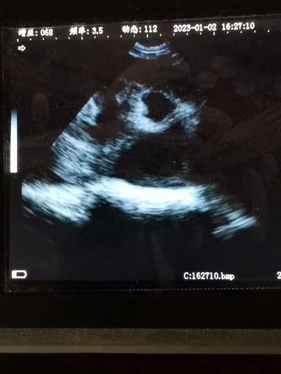
When using the DAWEI S1 portable veterinary ultrasound for examination in cattle, a 5.0 MHz or 7.5 MHz probe is inserted into the rectum. The earliest detection of the embryonic vesicle varies depending on the frequency of the probe. Using a 5.0 MHz probe, the embryonic vesicle can be detected around 12 to 14 days after mating, while a 7.5 MHz probe can detect the fetal vesicle as early as 9 days after mating. Fetal structures can be observed around 13 days after mating, and fetal heartbeat can be observed from day 22.
As a handheld portable veterinary ultrasound machine, DAWEI S1-VET has the advantages of light weight, easy to carry, long running time, high-definition image display, easy and fast early pregnancy monitoring.
Post time: Jun-25-2023