


Why equine limb imaging is harder than it looks
Equine limb radiography is often performed for lameness workups, traumatic injuries, follow-ups, and pre‑purchase exams (PPE). Compared with many small‑animal studies, it’s less forgiving because you’re balancing:
- Fine bony detail: crisp cortical margins, subtle joint changes, small fragments.
- Speed: horses don’t hold perfect positioning for long—slow workflow increases motion and retakes.
- Field conditions: barns, stables, uneven flooring, dust, variable power.
- Safety and dose management: fewer repeats means less exposure and less time in risky positions.
Think “system,” not just “X‑ray power”: the 4 building blocks
Many buyers focus on generator power alone. For equine limb radiography, overall results depend on the entire imaging chain:
- X‑ray generator: stable output, predictable technique settings, practical portability for your environment.
- Wireless flat panel detector (FPD): size, durability, wireless reliability, and image availability speed.
- Stand / positioning solution: how quickly you can get repeatable angles and lock them in.
- DR software + workflow (DICOM/PACS): image processing, protocol presets (APR), storage, transfer, and reporting.
A classic overview from dvm360 highlights that DR’s major workflow advantage is near‑instant image availability—helping teams spot positioning errors earlier and reduce downtime compared with slower processing workflows (dvm360, Digital radiography equipment: Exploring your options, 2009).
How to choose an equine DR system: 8 decision factors that matter most
1) Detector size (and the useful imaging area)
Equine limb work commonly includes the hoof, pastern, fetlock, and carpus.
- A large-format wireless flat panel detector makes it easier to cover multiple views quickly—especially in field conditions.
- Don’t look only at the outside dimensions; ask about the active imaging area and how it affects your common projections.
2) Image speed: preview time and field rhythm
In the field, your “good positioning window” is short. You want:
- Fast preview to confirm centering, rotation, and collimation.
- Stable wireless transfer that doesn’t become the bottleneck.
Practical takeaway: if you do PPE sets or multi-view series, slow images don’t just slow imaging—they slow the entire appointment.
3) Dose vs. diagnostic quality (and why processing matters)
“Low dose” is often marketed, but the true goal is diagnostic-quality images at a reasonable dose.
- Consistent image processing (contrast, noise management, edge detail) often determines whether subtle lesions are readable.
- As dvm360 notes, differences in final image appearance frequently reflect vendor image processing workflow, not just hardware class.
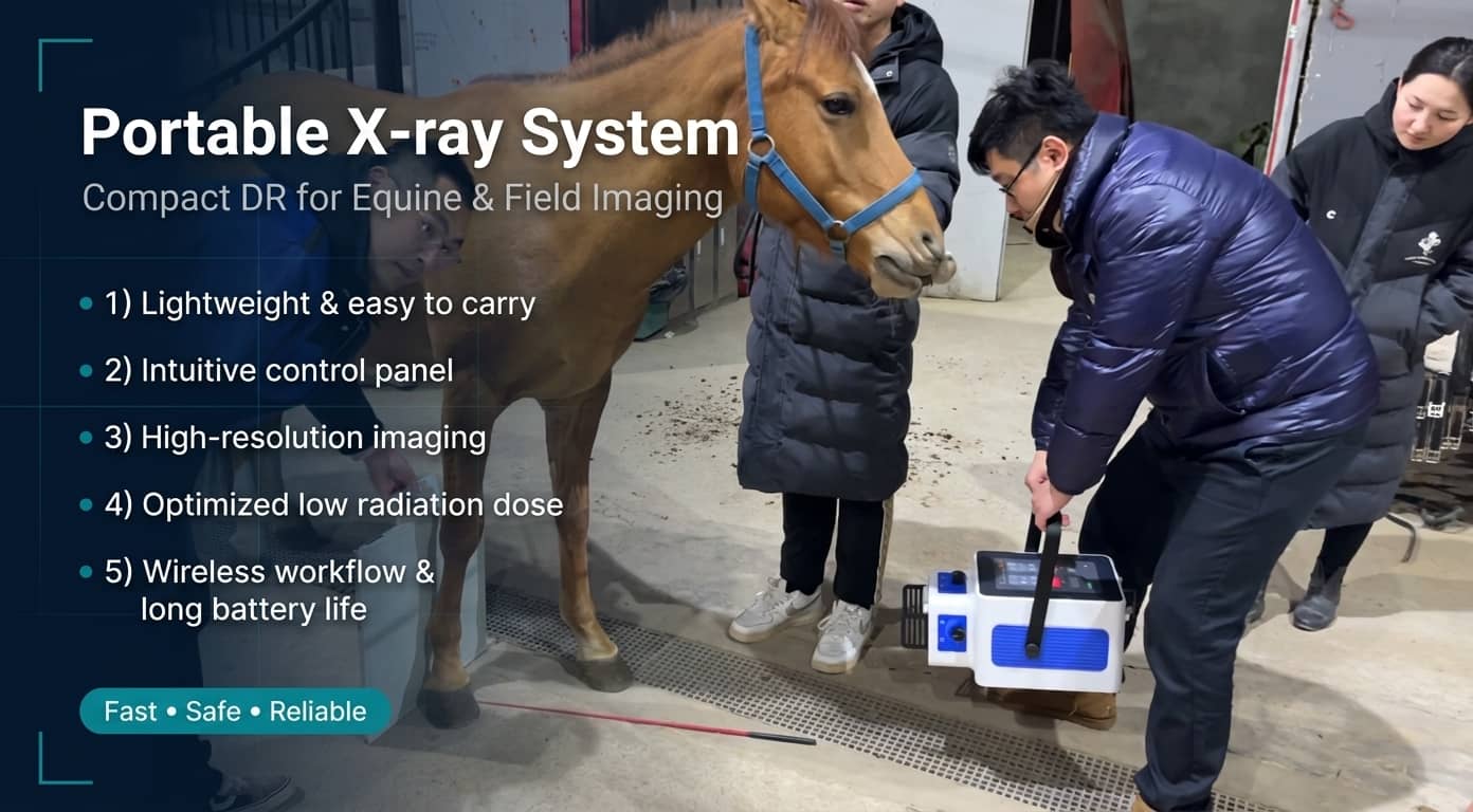
- Portable veterinary X‑ray / portable equine DR: ideal for mobile practice and stable calls; prioritize fast setup, portability, and power flexibility.
- Stationary large animal X‑ray: ideal for clinics and imaging centers; prioritize throughput and repeatability.
4) Portable vs. stationary: match the generator to your workflow
A simple rule: decide whether you need “radiography anywhere” or “maximum efficiency in one optimized room.”
5) Positioning and standard views: equipment should support your technique
For the fetlock, IMV Imaging describes four standard projections:
- Lateromedial (LM)
- Dorsopalmar(/plantar) (DP)
- Dorsolateral–palmaromedial oblique (DLPMO)
- Dorsomedial–palmarolateral oblique (DMPLO)
They also emphasize practical points that reduce repeats: clean the limb to avoid artefacts, consider sedation to improve safety and efficiency, and aim for even weight bearing with a vertical limb (IMV Imaging, Radiography techniques of the equine fetlock joint, 2019).
So on the equipment side, check whether:
- the stand locks quickly and holds angles reliably
- the panel placement is stable for hoof-level work
- the detector is built for real stable conditions (dust, knocks, transport)
6) DR software, APR presets, and DICOM/PACS integration
Equine limb radiography is rarely “one picture.” A smooth workflow is usually:
Registration → anatomical selection → protocol/preset (APR) → acquisition → processing → export/archive/print.
Look for:
- a veterinary DR software workflow that’s fast and learnable
- flexible APR protocol libraries you can customize
- solid DICOM support for PACS/RIS and referral sharing
7) Reliability in barns: durability + service response
Stable environments punish equipment: dust, humidity, vibration, accidental impacts.
- Detector ruggedness and protective design matter.
- Equally important: training, warranty, spare parts, and service turnaround.
8) Choose by scenario: mobile team vs. imaging center
Use this as a quick match:
- Mobile equine vet / field service: portable generator + wireless FPD + lightweight stand + laptop workstation
- Equine clinic (mid-volume): portable system for most calls + a dedicated room/upgrade path for complex cases
- High-throughput imaging center: stationary system + strong stand/positioning efficiency + PACS-first workflow
“Best DR systems” isn’t one model—it’s 3 high-converting setups
Setup A: Portable equine DR (mobile practice / stable calls)
What to prioritize:
- stable, practical portable generator workflow (fast setup, consistent output)
- large-format wireless FPD to reduce repositioning stress
- lightweight, foldable stand
- fast image processing + DICOM-ready software
Setup B: General-purpose large animal DR (clinic + hospitalized cases)
What to prioritize:
- a workflow that scales with volume (less clicking, fewer manual steps)
- repeatable stand positioning
- DICOM-first image sharing and archiving
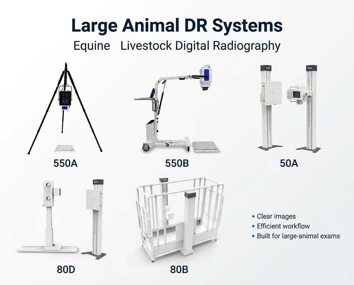
Setup C: Stationary, high-throughput large animal X-ray DR (imaging center)
What to prioritize:
- high-throughput workflow and consistent positioning
- motorized lift / tracking features (reduce waiting and reposition time)
- animal-focused DR software for stable output and fewer repeats
Product internal-link anchor example (copy/paste):RV-80D Stationary Large Animal DR System
FAQ
Do I really need a wireless flat panel detector for equine limbs?
Not always—but for mobile and stable settings, wireless FPDs typically improve speed and flexibility. The deciding factors are wireless stability, durability, and how fast you can confirm positioning.
How do I reduce retakes on hoof/fetlock radiographs?
Across training resources, three practical steps show up repeatedly:
- keep the limb/hoof clean to avoid artefacts
- consider sedation when appropriate to reduce motion and risk
- insist on consistent, standard views and even weight bearing
Equipment supports this by making positioning faster and images available sooner.
What DR software features matter most in equine practice?
- APR presets (fewer technique errors)
- strong processing and measurement tools
DICOM/PACS connectivity (referrals, archives, remote consults
Post time: Apr-20-2026