


In modern livestock production, the gastrointestinal (GI) health of cattle and sheep is crucial to their performance and overall welfare. GI diseases not only result in economic losses but also cause considerable animal suffering. As a veterinary ultrasound manufacturer, we recognize the vital role advanced diagnostic technologies play in the early detection and accurate assessment of gastrointestinal problems. This article explores common GI issues in cattle and sheep, the application of ultrasound technology in gastrointestinal scanning, relevant case studies, and global veterinary trends in GI diagnostics.
1. Common Gastrointestinal Issues in Livestock
Gastrointestinal diseases are among the most frequently encountered health problems in cattle and sheep, caused by various factors including nutrition, infections, parasites, and improper management.
(1) Common GI Issues in Cattle
Cattle have a complex digestive system, particularly as ruminants, and are highly sensitive to various stressors. Common GI conditions include:
Rumen Acidosis: Caused by high-concentrate diets or sudden feed changes, leading to a drop in rumen pH, poor digestion, reduced feed intake, and potentially laminitis or death.
Traumatic Reticulitis/Pericarditis (“Hardware Disease”): Occurs when cattle ingest metallic foreign bodies (e.g., nails, wires) that penetrate the reticulum wall, possibly causing localized inflammation or pericarditis.
Displaced Abomasum (DA): Often seen in postpartum dairy cows. The abomasum moves to the left (LDA) or right (RDA) side of the abdomen, disrupting digestion and significantly reducing feed intake and milk production.
Intestinal Obstruction: Caused by foreign bodies, volvulus, intussusception, or tumors, leading to severe abdominal pain, vomiting (rare in cattle), and difficulty passing feces.
Paratuberculosis (Johne’s Disease): A chronic and progressive intestinal disease caused by Mycobacterium avium subsp. paratuberculosis, characterized by persistent diarrhea, weight loss, and reduced productivity.
Gastrointestinal Parasitism: Caused by nematodes, tapeworms, or coccidia, leading to malabsorption, diarrhea, anemia, and stunted growth.
(2) Common GI Issues in Sheep
While similar to cattle, sheep also suffer from specific gastrointestinal conditions:
Gastrointestinal Nematodiasis: Caused by parasites such as Haemonchus contortus and Ostertagia spp., common in sheep farming, resulting in anemia, edema, diarrhea, and weight loss.
Rumen Bloat: Can be primary (frothy) or secondary (free gas), due to gas accumulation in the rumen. It causes abdominal distension and may lead to respiratory distress or death.
Bacterial Enteritis in Lambs: Caused by E. coli or Salmonella, leading to diarrhea, dehydration, and high mortality rates.
Coccidiosis: Affects lambs and young sheep, causing watery or bloody diarrhea, emaciation, and poor growth.
Endotoxemia: Secondary to severe GI infections or digestive disturbances, where bacterial endotoxins enter the bloodstream, causing systemic inflammation.
2. How Veterianry Ultrasound Is Used to Scan the GI Tract
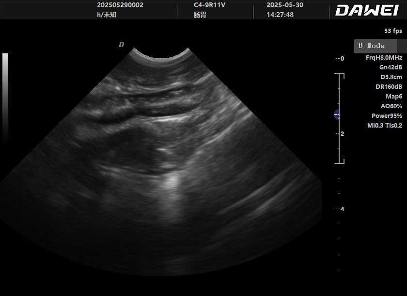
Ultrasound is a non-invasive, real-time, repeatable diagnostic tool that plays an increasingly vital role in identifying GI diseases in cattle and sheep. It utilizes high-frequency sound waves to produce images based on reflected echoes from internal structures.
Key Ultrasound Scanning Techniques and Considerations:
Probe Selection and Frequency:
Low-frequency convex probes (2.0–5.0 MHz): Ideal for deep tissue scanning, including the rumen, reticulum, abomasum, and intestines, with high penetration capabilities.
High-frequency linear probes (7.5–12.0 MHz): Suitable for superficial structures and detailed assessments, such as intestinal wall thickness, layer definition, and lymph nodes.
Animal Positioning and Restraint:
Animals are usually scanned in a standing position and may require sedation. The scanning area should be clipped and gel applied to ensure good contact and eliminate air bubbles.
Systematic Scanning Approach:
Cattle:
Left Abdomen: Evaluates the rumen and left-displaced abomasum. A normal rumen has a thick wall with coarse or liquid content. LDA appears as a crescent or oval structure filled with gas and fluid, often with a “ping” sound on percussion.
Right Abdomen: Evaluates the abomasum, duodenum, jejunum, ileum, cecum, and colon. RDA appears as an enlarged abomasum with abnormal gas content. Bowel dilation, wall thickening, loss of layer definition, and reduced motility are significant findings.
Xiphoid Region: Important for reticulum evaluation, especially in diagnosing traumatic reticulitis. Findings may include thickened reticulum wall, exudates, fibrin deposits, or visible foreign bodies.
Sheep:
The scanning approach is similar, but due to smaller body size, the entire GI tract is more accessible. Special attention should be given to intestinal wall thickness, motility, content, and lymph nodes.
Key Diagnostic Indicators:
Intestinal Wall Thickness: Normal walls are thin and regular; thickening suggests inflammation, edema, tumors, or lymphangiectasia.
Wall Layer Structure: Healthy intestines display five distinguishable layers. Loss of this structure indicates inflammation or infiltration.
Bowel Contents: Typically fluid, gas, or mixed. Abnormal contents (excess fluid, foreign bodies, fecaliths) may suggest obstruction.
Peristalsis: Normal intestines show regular motility. Hyperactivity may indicate early inflammation; hypomotility suggests ileus or obstruction.
Lymph Nodes: Enlarged mesenteric lymph nodes suggest infection or conditions like paratuberculosis.
Surrounding Structures: Look for free fluid, fibrin, or abscesses in the abdominal cavity.
3. Ultrasound Case Studies
Ultrasound offers veterinarians clear visualization of GI pathologies for accurate and timely diagnoses.
(1) Cattle Case Studies
Case 1: Left Displaced Abomasum (LDA)
A postpartum dairy cow presented with reduced appetite, sudden drop in milk yield, and left abdominal distension. Percussion and palpation suggested LDA. Ultrasound revealed a crescent-shaped structure with gas and fluid between the 9th–12th left intercostal spaces, confirming LDA. This guided surgical correction.
Case 2: Traumatic Reticulitis
A beef cow showed depression, poor feed intake, arched back, and decreased defecation. Palpation revealed xiphoid tenderness. Ultrasound showed thickened reticulum wall, localized exudate, and hyperechoic areas indicating potential foreign body penetration—classic signs of hardware disease.
(2) Sheep Case Studies
Case 1: Intestinal Lymphoma
An adult ewe suffered chronic weight loss and diarrhea. Abdominal palpation revealed a mass. Ultrasound showed a thickened small intestine wall with loss of normal layers and hypoechoic mass-like appearance, with enlarged mesenteric lymph nodes. Biopsy confirmed lymphoma. Ultrasound was key in assessing tumor extent and metastasis.
Case 2: Severe GI Parasitism in Lambs
A group of lambs displayed weight loss, edema, rough coats, and anemia. Ultrasound showed mildly thickened intestines, disrupted contents, and reduced motility. Although parasites weren’t directly visualized, ultrasound suggested inflammation consistent with parasitism, later confirmed via fecal testing.
4. International Focus on GI Diagnostics
Countries with advanced livestock industries—like Australia, the UK, and Brazil—integrate imaging, clinical signs, and lab diagnostics to address GI diseases.
Australia:
Emphasizes precise parasite control due to extensive pasture systems. Ultrasound helps assess bowel inflammation severity to guide deworming. Paratuberculosis control is also key, with ultrasound used to evaluate lymphadenopathy alongside serology and fecal culture.
United Kingdom:
Focuses on high-yield dairy cows and GI/metabolic diseases. Early detection and surgical planning for displaced abomasum heavily rely on ultrasound. Conditions like BVD and viral enteritis are also assessed through GI ultrasonography to aid differential diagnosis.
Brazil:
As a major beef producer, Brazil prioritizes growth performance and disease prevention. Nutritional GI disorders (e.g., acidosis) and parasitism are common. Ultrasound aids in distinguishing obstruction, tumors, and inflammation. In calves, it helps evaluate diarrheal severity and dehydration for fluid therapy planning.
Post time: Jul-08-2025