


Ultrasound technology has been widely used in livestock production, especially in veterinary medicine and animal husbandry.Animal Ultrasound (B-scan ultrasonography) is a non-invasive examination technique that utilizes the principle of ultrasonic imaging to observe and diagnose the internal organs and tissues of animals.
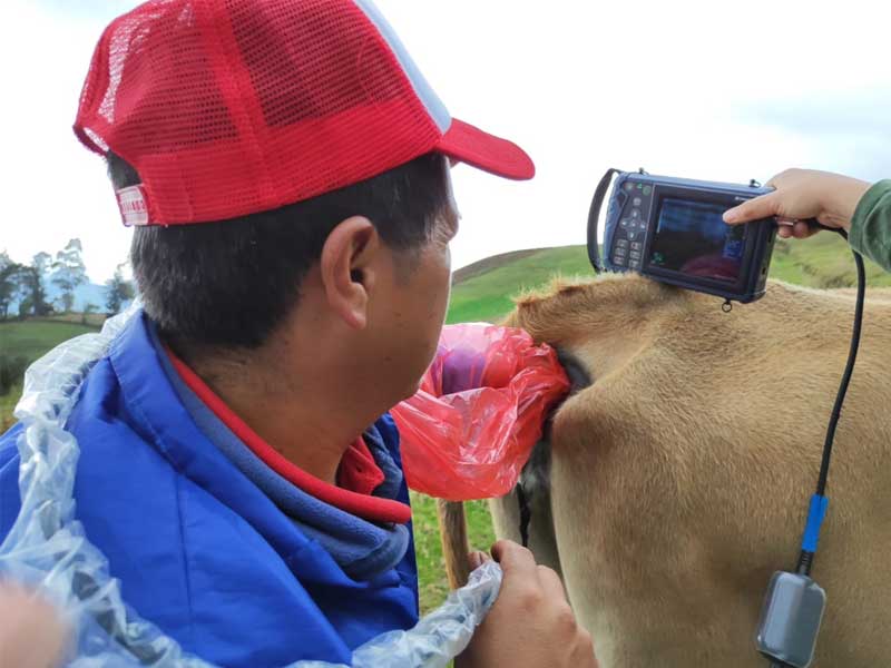
B-scan ultrasonography is a non-invasive technique that uses ultrasound imaging to visualize and diagnose internal organs and tissues of animals. Ultrasound pregnancy testing is a common method of determining whether a female animal (e.g., cow, sheep, pig, etc.) is pregnant, as well as the ability to identify empty females (e.g., cows, sheep, swine, etc.) as early as possible; at the same time, ultrasound of an animal’s abdomen allows veterinarians to confirm pregnancy and assess the number and developmental status of fetuses. Pregnancy monitoring is a crucial part of livestock production, which directly affects the reproductive efficiency and productivity of livestock. Using ultrasound technology, non-invasive testing can be performed in early pregnancy, and parameters such as fetal heartbeat, placenta, and fetal size can be observed, so that targeted management measures can be taken to improve reproductive efficiency.
Determination of backfat thickness and calculation of eye muscle area in female animals. Backfat thickness is the thickness of subcutaneous fat on the back of the animal. Backfat thickness is widely used in livestock production to assess body fat content and body mass of animals, especially for breeding and feeding management of meat animals such as pigs, cattle and sheep. Evaluating an animal’s body weight, assessing meat quality, and monitoring backfat thickness through ultrasound measurements of backfat thickness can help determine an animal’s feeding program to ensure that the animal is between appropriate levels of fatness and leanness, which can help to improve productivity and meat quality.Ocular muscle area refers to the cross-sectional area of the ocular muscles within the body of an animal. In livestock production, oculomotor area is commonly used to assess the leanness and meat quality of meat animals, oculomotor area is directly correlated with the leanness of the animal, with larger oculomotor area usually meaning more lean meat, which is important for producing high quality meat animals.
Determination of equine tendon ligaments, Tendons and ligaments are important structures that connect muscle to bone and play a key role in the movement and support of the animal. Therefore, the health of tendons and ligaments is closely related to an animal’s muscle mass and athletic performance. Detection of tendon ligaments by ultrasound can indirectly assess the muscle quality and functional status of animals, which provides an important reference for selection and breeding management. Through ultrasound examination of tendon ligaments, breeders and veterinarians can detect potential sports injuries, such as tendon strain or ligament strain, so that timely treatment measures can be taken to prevent further deterioration of the injury.
Animal ultrasound technology helps to examine the thoracic and abdominal cavities of animals to detect abnormalities in internal organs such as the heart, lungs, liver, spleen, etc. Ultrasound can help veterinarians to detect organ injuries and lesions caused by diseases. For example, ultrasound can detect lung infections, fluid buildup, or other pathological changes when examining an animal’s lungs. When examining the abdominal cavity, it can detect liver lesions, enlarged spleens, kidney stones, etc. Ultrasound also has important applications in the reproductive management of animals. Ultrasound examination of the uterus and ovaries of the female animal can determine the animal’s physiological cycle and ovulation, which can help determine the right time for breeding.
Post time: Jul-31-2023