


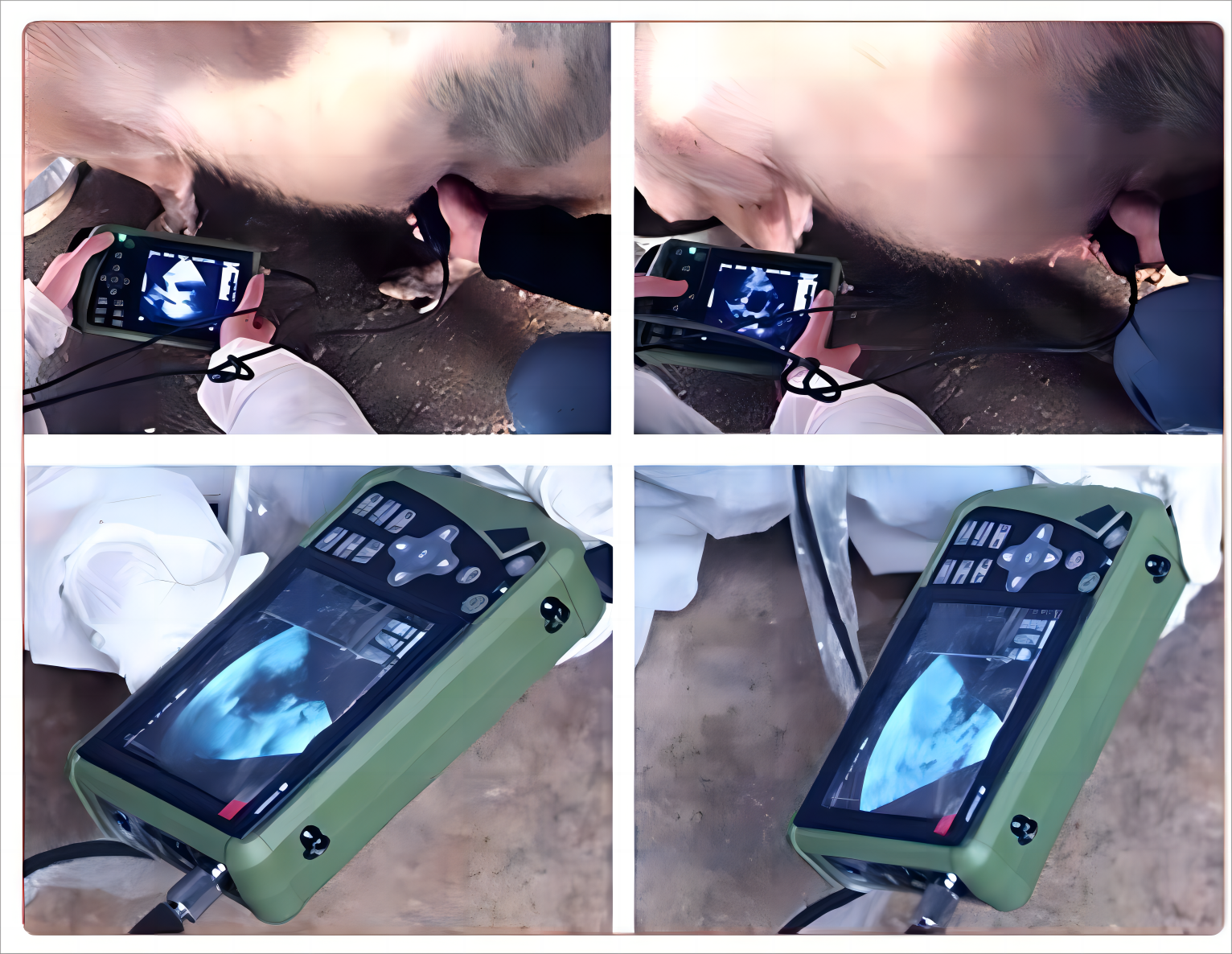
The advent of portable swine ultrasound machines will greatly facilitate reproductive imaging of sows and may assist in making management decisions, which can be based on an accurate diagnosis of the reproductive state of a reserve sow or gilt at any given time.
It is at this time that piglets form and grow inside the sow.
Typically, the length of a sow’s pregnancy or gestation is about 115 days, during which time the piglet goes through different developmental stages, which we have categorized into 5 main phases: days 0-15, days 13-30, days 30-77, days 77-90, and days 90-114.
Below are the characteristics of the ultrasound images of each stage detected using a swine ultrasound machine: Characteristics of ultrasound images of sows at days 0-15 of pregnancy detected by a swine ultrasound machine: The first sign of estrus is considered to be day 0 of the estrous cycle, ovulation occurs in the first 24-48 hours after estrus begins, the fertilized embryo elongates and migrates to find space in the uterus, and during this period sow fights and traumas often have little or no effect on the survival of embryos, as embryos are often not able to survive because of their lack of ability to survive in the uterus. survival because the embryo has not yet formed an attachment to the uterine wall, and any loss of embryos at this time is usually influenced by disruption of preovulatory oocyte quality, sperm quality, or hormonal signaling prior to fertilization;
Attachment of the embryo to the uterus occurs around days 12-15: in pigs, four or more viable embryos must establish attachment to the uterine wall for a pregnancy to be viable and continue beyond this point, during the attachment stage, the embryos begin to line up along the uterine horns, and if there are enough viable embryos attached, the pregnancy will be recognized on day 11-12, and embryos that fail to attach to the uterine wall will not be able to embryos that fail to attach to the uterine wall will not survive the pregnancy. If there are not enough embryos attached to the uterine wall (less than 4), the pregnancy will not be recognized and the sow will return to estrus on day 21 after breeding. If the embryos are successfully attached to the uterine wall, they will begin to form in their own placenta because each piglet has its own placenta. Pigs have an epithelial placenta, which means that the placenta does not invade the uterine tissues as much as other types of placenta that fit snugly against the uterine wall and form a groove for attachment, usually compared to Velcro, where each piglet has its own placenta, separating each fetus from the rest of the litter away and prevents the loss of one fetus from affecting the survival of the other piglets in the litter. However, this also means that each placenta needs to be fully attached to the uterus, and a lack of space can affect placental development in any pig. This means that the larger the bedding, the less room there is for the placenta to attach, which can result in smaller piglets.
Characteristics of the ultrasound image of a pig ultrasound machine for detecting sows on days 13-30 of pregnancy: In this timeframe, the initial placental dilatation, which occurs fairly quickly, occurs between days 27-40, and around day 30, which is in the middle of the dilatation phase, the sows can be checked ultrasonographically to see if they are pregnant. When using a veterinary ultrasound machine, a fluid-filled sac indicates normal mat development.
In group gestation penning systems, if sows are not penned immediately after breeding, it is best to wait until pregnancy is confirmed before penning sows together, by which time placental attachment is considered sufficient to survive the fighting that occurs between sows.
Characteristics of ultrasound images of sows detected by a swine ultrasound machine on days 30-77 of pregnancy: During this period, visible organ development begins and bones start to calcify on days 35-45. Sometimes, for various reasons, individual fetal pigs stop developing and die in utero.
Fetal pigs that die during gestation may result in mummification at delivery. Mummification is the death of the fetus after calcification of the skeleton and therefore cannot be reabsorbed. Instead, it breaks down and mummifies in the placenta
For hog producers, the discovery of many mummified fetuses in the bedding at farrowing may be a sign that the sow has been traumatized early in gestation or by her developing offspring, and trauma may include rough handling, poor nutrition, environmental stress, or disease stress.
Porcine ultrasound machine detects ultrasound images of sows on days 77-90 of pregnancy characterizing eventual placental dilatation beginning on day 77.
This is an intrauterine marker of late gestation, but late gestation is observed externally in the sow through visible mammary tissue expansion.
At this point, colostrum and milk production begin in addition to continued fetal growth.
Post time: Jan-02-2024