


In modern scale pig farms, sow reproduction disorders are a common phenomenon, there are many reasons that can cause sow disorders, such as: inbreeding genetics, loss of feed nutrients, environmental stress, bacterial infections, viral infections, etc., which cause a lot of economic losses to the farms, and some sows give birth to sows prematurely, produce stillbirths, miscarriages, embryo resorption, and some sows suffer from endometritis, uterine storage of pus, and some sows have been mated for a long time and are infertile. Infertile, easy to cause ineffective feeding of sows and premature elimination of sows, more and more pig farm bosses use veterinary ultrasound machine to find out sow reproduction disorders in advance, timely treatment of sow reproduction diseases and find empty womb phenomenon.
1 Ultrasound and veterinary ultrasound
Our human ears can hear the sound wave frequency of 20Hz to 20kHz, when the frequency of sound wave is higher than 20kHz we can’t hear it, and we call this frequency higher than 20kHz ultrasonic wave. Ultrasonic waves have good directionality, strong penetrating ability, easy to obtain a more concentrated sound energy, especially in the water long-distance propagation. In human medicine, the use of ultrasound’s physical properties to analyze and diagnose diseases produce ultrasound, ultrasound is to transmit ultrasound waves to the human body, while accepting the body’s internal organs of the reflected waves, the image information obtained will be reflected on the screen. China began to use simple ultrasound to observe the human liver in 1947, when the image was black-and-white and fuzzy, and was initially used to detect fetal tissue structure and measure how big and long the fetus was.In the 1980s, red-blue ultrasound based on black-and-white ultrasound was used to observe fetal blood flow, but black-and-white ultrasound and red-blue ultrasound can only observe the internal organs of the human body from one direction. With the rapid development of computers, the emergence of three-dimensional ultrasound, can be viewed from multiple directions through the screen to observe the internal organs of the human body, to obtain the image is three-dimensional, more comprehensive, real and clear. With the development of imaging and printing industry, the emergence of four-dimensional color ultrasound, can print color photos, more comprehensive, real and clear understanding of the internal organs of the human body. Many people use ultrasound in human medicine to analyze and diagnose animal diseases, resulting in veterinary ultrasound. Veterinary ultrasound is a simplification of human ultrasound, which is easy to operate. Veterinary ultrasound is to transmit ultrasound to the animal, while accepting the animal internal organs of the reflected wave, the image information obtained will be reflected on the screen
2.The working principle of animal ultrasound
Animal ultrasound contains the launching, scanning, receiving, signal processing and screen display, that is, divided into two parts of the host and the probe. Probe can be installed 1 pressure wafer or more pressure wafer, the wafer in turn work, can transmit and receive sound energy, become a transducer. Probe according to the frequency classification, there are single-frequency, multi-frequency and broadband and other types. The probe uses the ultrasonic waves sent from the host, generally with anastomosis agent coated on the probe to better contact with the animal surface, ultrasonic waves in two different tissues at the interface to produce reflection, refraction, scattering, bypassing, reduction and the sound source and receiver relative motion to produce Doppler and other physical characteristics, according to monitoring the delay time of their echoes, the strength of which can be determined by the distance of the organ and the nature of the. After the electronic circuit and computer processing, the physical characteristics are collected and displayed on the screen through the host computer. It becomes different images. The key component of ultrasound is what we call ultrasound probe, which has a set of internal ultrasound transducer, is made of a group of special crystals with piezoelectric effect. This piezoelectric crystal has a special property, that is, in the specific direction of the crystal with a voltage, the crystal will be deformed, and in turn when the crystal is deformed, the corresponding direction of the voltage will be generated to achieve the conversion of electrical signals and ultrasound.
3 Veterinary ultrasound machine in the sow reproduction disorders of practical application
3.1 Veterinary ultrasound machine can monitor follicle development and ovulation, providing reliable scientific basis for when to breed and improve breeding rate.
Sow is a perennial multiple estrus animal, as long as the sexual maturity, generally every 18~23 days estrus, no mating, will be repeated estrus. The ovaries of sows also show cyclic changes with the estrous cycle. Two to three days before the onset of estrus in sows, follicles begin to enlarge rapidly until 18h after estrus. The follicles were inconsistent in size, and mature follicles were mussel-red in color. In sows, “hemorrhagic follicles” were often found due to blood infiltration into the follicular cavity from arterial congestion. When a transparent area appears at the top of the follicle, it indicates that ovulation is imminent. The corpus luteum is fully formed on the 6th to 8th day of the estrous cycle, and its secretory action is maintained until the 16th day, and then it rapidly degenerates. The corpus luteum of the sow is dark red at the beginning because the lumen is filled with dark red coagulated blood clots, and gradually changes to light purple by the 15th day of the estrous cycle, to light yellow by the 18th day, and to white later (see Figure 1).
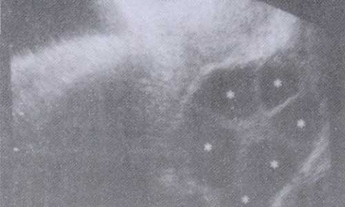
FIG.1 Maturation stage of sow follicles
3.2 Early pregnancy monitoring by veterinary ultrasound machines can detect empty sows as early as 18 days after mating in time for early treatment accordingly.

FIG. 2 The sow is not pregnant
If the sow is not pregnant, there is no gestational sac, and the veterinary ultrasound will show a faint white cloud (see Figure 2), while there is amniotic fluid and a small gestational sac is formed 18 days after mating, and 25 to 30 days after mating is the best time to monitor, at this time, the gestational sac looks like an obvious and regular round black circle, and the regular round black circle represents the number of heads of the pig (see Figure 3). Amniotic fluid can be observed around 21 to 35 days of pregnancy, the amniotic fluid image is like a honeycomb, the black circle is getting bigger and bigger, the amniotic fluid is getting less and less, and the fetus of the pig is like a black ball of skin floating in the air at 30 to 35 days. The uterus of no pregnant sows has no black circle around 25~60 days of breeding and will show a regular flat white cloud-like shape.
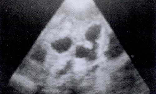
FIG.3 Sow at 25 days of pregnancy
3.3 Ultrasound monitoring during pregnancy can detect stillbirths, abortions, embryo resorption, etc., as well as estimate the number of litters carried. Sow is prone to hidden abortion after 20 days of breeding, and it is measured in more than 20 days when the sow is already pregnant, but later it does not give birth, the gestational sac is absorbed by the uterus, and it does not show the symptoms of abortion, so we have to carry out the 2nd pregnancy test after 35~45 days of breeding to accurately master the pregnancy of the sow, and the sow will have the symptoms of abortion if it has aborted after 40 days of breeding, and we can observe it in time with our careful attention. If there is no abortion, if the ultrasound detects that the sow is already pregnant, it can show that the sow is already pregnant.
3.4 Ultrasound monitoring during parturition can determine fetal viability and whether the fetus and fetal coat are exhausted. Images after 70 days of pregnancy, piglet bones have been calcified, amniotic fluid is absorbed, at this time there is no longer a black gestational sac, there is no round black circle on the image, the performance of an arc like a dotted line piglet vertebrae (see Figure 4), images after 90 days of pregnancy, we can see the piglet’s fetal heart in the beating, there is no fetal heart in the beating indicates that the fetus is dead. Veterinary ultrasound monitoring allows us to observe the recovery of the uterus of the sow after parturition, and to diagnose reproductive disorders such as endometritis, uterine pus accumulation, and fluid retention. Sows with endometritis will have pus accumulation and fluid retention, which is reflected in the veterinary ultrasound image as a torn cotton mattress that is irregular and uneven (see Figure 5).
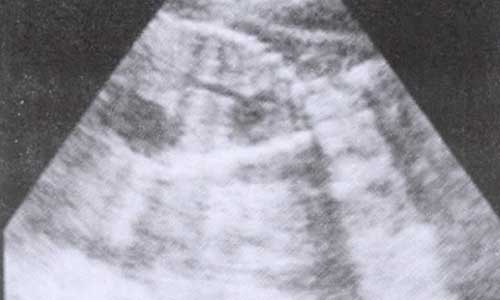
FIG. 4 Sow 75 days pregnant
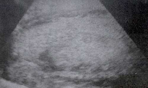
FIG. 5 Sow with endometritis with pus accumulation
With the development of the times, the progress of science and technology, animal husbandry is also changing rapidly, the traditional pig farms are no longer in line with the needs of today’s society, technological pig farming has become an inevitable trend in the farming industry, modern pig farms in all aspects of the equipment is also higher and higher, more and more pig farms feel the use of veterinary ultrasound, began to consider the purchase of veterinary ultrasound, but some people believe that the small size of the farms are not suitable for the use of Veterinary ultrasound, this idea is wrong, because scientific equipment can provide data in all aspects, improve efficiency, timely treatment of sow reproductive diseases and find empty pregnancy phenomenon, to bring more and better economic benefits.
Post time: Aug-22-2023